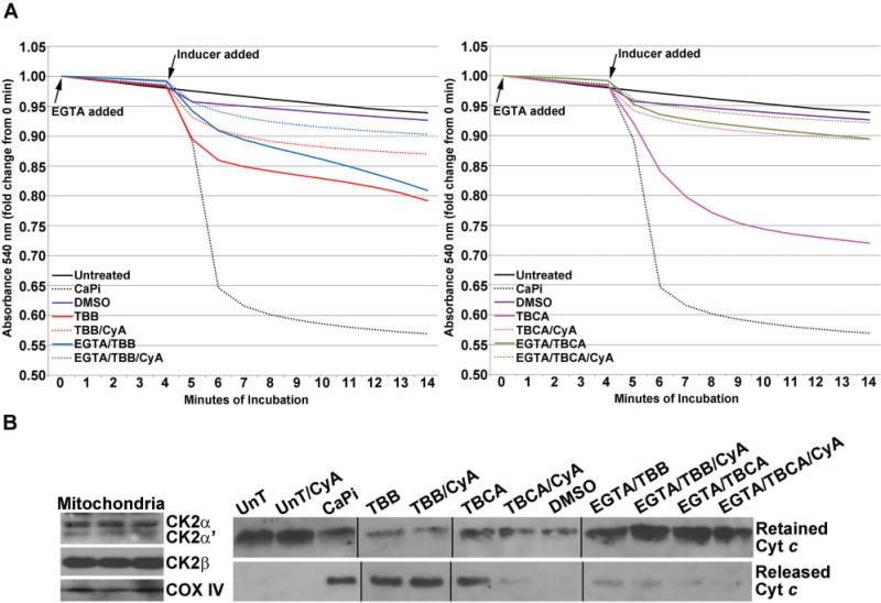
Fig. 5. Effect of CK2 inhibitors on membrane permeability transition in isolated mitochondria.

(A) Effect of TBB (left panel) or TBCA (right panel) treatment on mitochondrial permeability transition. Purified mitochondria were subjected to various treatments as shown in the graph legends. Mitochondrial respiration was maintained using 10 mM Na-succinate as the substrate. When used, EGTA or cyclosporin A was added at time zero; “inducers” were added at the start of the 5th min after mitochondria equilibration in the incubation medium and baseline absorbance (540 nm) had been captured for 4 min. Absorbance was measured for an additional 10 min following the addition of the various chemicals as indicated. The concentrations of various agents were: 80 μM TBB; 80 μM TBCA; 70 μM Ca2+, 3 mM Pi; 0.5 μM cyclosporin A; 150 μM EGTA. A volume of DMSO equivalent to the volume of TBB/TBCA was used. All other details are as described under Materials and Methods. (B) Left panel, rat CK2α, CK2α′, CK2β′ and COX IV were detected in all preparations of rat liver mitochondria by western blot analysis; 3 representative preparations are shown. Right panel, cytochrome c that was retained in the mitochondria or released in the medium corresponding to the various treatments under A (left and right panels) was detected by western blot analysis.
