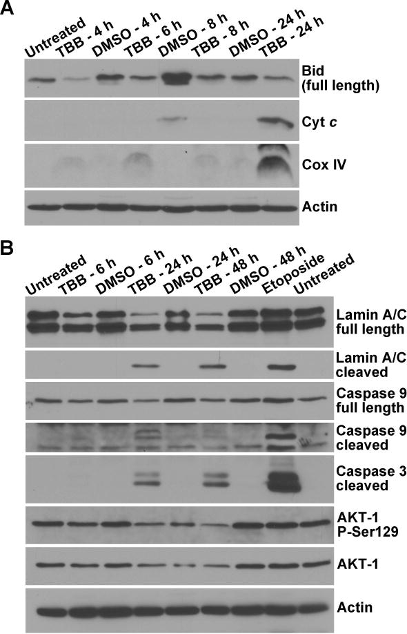
Fig. 6. Temporal response of CK2 substrate and apoptotic signals following TBB mediated inhibition of CK2 in PC3-LN4 cells.

(A) Effect of 80 μM TBB treatment of cells for varying times on apoptotic signals measured in the cytosol. Cells were treated with TBB for 4, 6, 8 and 24 h. Analysis of Bid, cytochrome c, and Cox IV was carried out in the purified mitochondria-free cytosolic fractions. (B) Effect of 80 μM TBB treatment of cells for varying times in whole cell lysates. Cells were treated with TBB for 6, 24, and 48 h as shown, with DMSO as the corresponding control. Full length and cleaved lamin A/C, full length and cleaved caspase 9, cleaved caspase 3, total and phospho-Ser129 AKT-1 were analyzed. Etoposide (100 μM for 24 h) was employed as the positive control for induction of apoptosis. All other details were as described under Materials and Methods.
