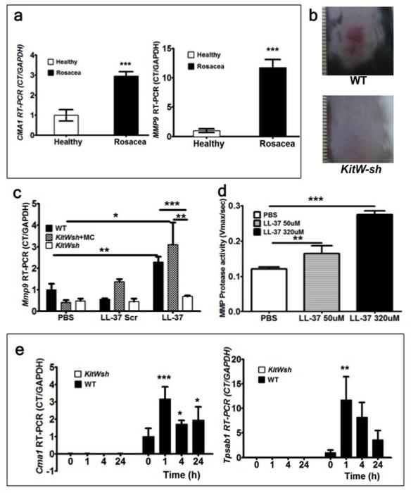
Figure 1. (a-e) MC proteases and MMP-9 are crucial for rosacea inflammation development.
(a) Human biopsy samples from healthy subjects (n=6) and rosacea subjects (n=6) were assessed for CMA1 and MMP9 mRNA expressions. (b) The back skins of wild type C57BL/6 (WT) and MC-deficient (KitW-sh) mice were injected intradermally with Cath LL-37. Pictures were taken 72 hours after LL-37 injections. (c) RT-PCR of Mmp9 mRNA expression in skin biopsies from WT, WT MC reconstituted KitW-sh (KitWsh) and KitW-sh. Mouse skin was injected with LL-37, LL-37 scrambled peptide (LL-37 Scr) or PBS control (PBS) and harvested after 72 hours of observation. (d) Assessment of MMP protease activity in WT mouse skin following challenge with PBS or Cath LL-37 at 50 and 320 μM. (e) mouse Chymase (Cma1) and mouse Tryptase (Tpsab1) were analyzed by RT-PCR in the skin from WT or KitW-sh mice (KitWsh) at 0, 1, 4, and 24 hrs after Cath LL-37 challenge. All of the experiments were repeated at least three times. Statistics: Mann Whitney test, one-way and two-way ANOVA*p<0.05, **p<0.01, ***p<0.001 (n=3)

