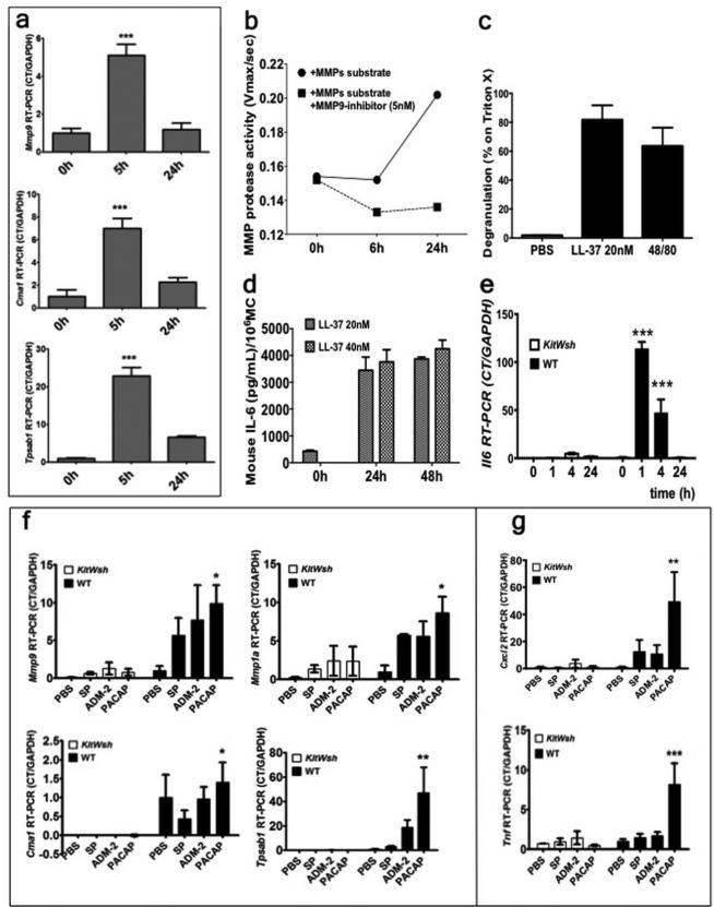
Figure 2. (a-e) Cath LL-37 induces pro-inflammatory responses.
(a) in vitro mMCs expression of Metallo protease 9 (Mmp9), Chymase (Cma1) and Tryptase (Tpsab1) after Cath LL-37 stimulation. (b) mMC Metallo pretease activity (MMP protease activity) after Cath LL-37 challenge. (c) mMCs degranulation (as β hexosaminidase release) in response to 24 hrs treatment with 20nM LL-37, positive control (48/80) and negative control (PBS). (d) ELISA IL-6 level (as pg/ml per million MCs) in mMC culture, 24 and 48 hours after 20 and 40 nM LL-37 treatment. (e) In vivo Il6 mRNA expression in mouse skin after 0, 1, 4, and 24 hrs of Cath LL-37 challenge in WT and MC deficient mice (KitWsh). (f-g) Neuropeptide-induced pro-inflammatory cytokine expression in skin is decreased in the absence of MCs in KitW-shmice. WT and KitW-sh (KitWsh), mice were injected with PBS or neuropeptide (SP, substance P; ADM-2, adrenomedullin-2; PACAP, pituitary adenylate cyclase-activating peptide) intradermally. (f) mRNA expressions of mouse Chymase (Cma1), mouse Tryptase (Tpsab1), mouse metalloprotease 9 (Mmp9), and metalloprotease 1 (Mmp1a) after NP challenge. (g) mRNA expressions of proinflammatory cytokines (Cxcl2 and Tnf) after NP challenge. Statistics: two-way ANOVA*p<0.05, **p<0.01, ***p<0.001 (n=3) repeated 3 times.

