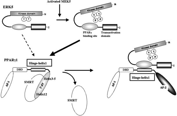
Figure 2. Model for the ERK5-PPARγ interaction-mediated PPARγ transactivation.
The position of Helix 12 is regulated by ligand binding. When the PPARγ ligand binds to the receptor, Helix 12 folds back to form a part of the co-activator binding surface, and inhibits corepressor (such as SMRT) binding to PPARγ94. The co-repressor interaction surface requires Helix 3-595. We found a critical role of the PPARγ hinge-helix 1 domain in ERK5-mediated PPARγ transactivation. The inactive N-terminus kinase domain of ERK5 inhibits its own transactivation and PPARγ binding. After ERK5 activation the inhibitory effect of the N-terminus domain decreases, and subsequently the middle region can fully interact with the hinge-helix 1 region of PPARγ. The association of ERK5 with the hinge-helix 1 region of PPARγ releases co-repressor of SMRT and induces full activation of PPARγ26. AF-1/2: Activating function (AF)-1/2 transactivation domain, DBD: DNA binding domain. Reprinted and modified from Akaike et al26.

