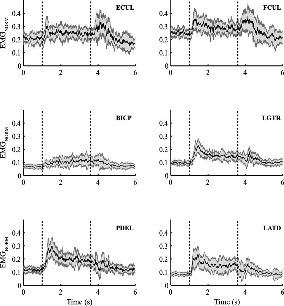
Figure 3.
EMG traces for a subset of muscles, extensor carpi ulnaris (ECUL), flexor carpi ulnaris (FCUL), biceps (BICP), triceps long head (LGTR), posterior deltoid (PDEL), and latissimus dorsi (LATD) for a representative subject (Subject #2) with TDWELL = 2 s. Averages across repetitive trials are presented; the shaded areas represent standard errors computed across trials. Muscle activations for each subject were normalized according to the averaged activity of the respective muscle during Position-holding trials (see Methods for details). The vertical dashed lines mark the start and the end of the perturbation force application.

