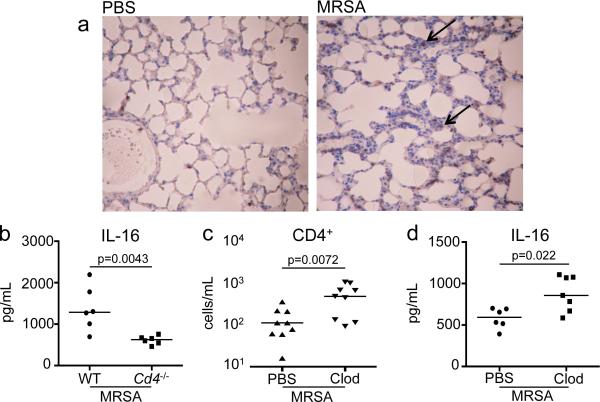
Figure 6. IL-16 is produced by CD4+ cells.
(a) Immunohistochemistry by standard methods with anti-IL-16 antibody on fixed whole lungs of C57BL/6J WT given PBS or infected with 107 MRSA for 24 hours. Magnification 100x. Black arrows point to areas of IL-16 staining (brown). (b) Age and sex-matched cohorts of C57BL/6J WT or Cd4−/− mice were inoculated intranasally with 107 MRSA and IL-16 was quantified in BALF collected by ELISA. Clodronate (Clod) or PBS liposome treated mice were given 107 MRSA for 24 hours. BALF stained for (c) CD4+ populations were analyzed by flow cytometry and (d) IL-16 was measured by ELISA. Each dot represents an individual mouse and data is combined from 2 separate experiments. Lines represent median values.

