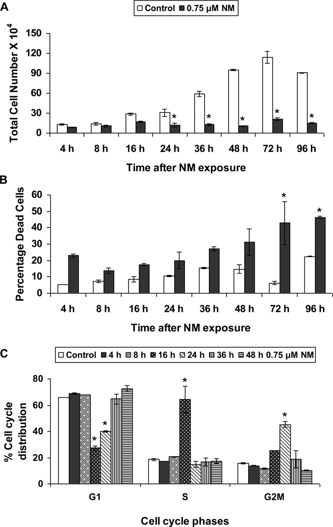
Fig. 1. Effect of NM exposure on cell proliferation and cell cycle progression in JB6 cells.
JB6 cells plated overnight in 60 mm plates were exposed to either DMSO or 0.75 µM NM for 4 to 96 h. At the end of the exposure times, all cells, including the floating dead cells were collected, subjected to Trypan blue exclusion assay using a hemocytometer for counting the cells. The total number of cells (a) and the percentage dead cells (b) were calculated as described under material and methods. Following similar exposures, FACS analysis was carried out to determine the cell cycle progression following NM exposure (c). Cells collected after 4 to 48 h of exposures were incubated overnight with saponin/PI at 4°C and subjected to FACS analysis as described in material and methods. Data are presented as mean ± SEM, n = 3. * P< 0.05 as compared to control.

