Fig. 1.
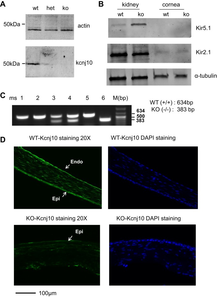
Kcnj10 is expressed in mouse cornea. A: Western blot was employed to detect the expression of Kcnj10 in mouse corneas dissected from Kcnj10−/− (KO), wild-type (WT), and Kcnj10+/− (Het) littermates, respectively. B: Western blot was used to examine the expression of Kir5.1 (top) and Kir2.1 (middle) in kidney and corneal tissue. C: mice used in the experiments were characterized by genotyping, 634 bp for WT, 383 bp for KO, and double bands for Het. D: fluorescent immunostaining images showed the expression of Kcnj10 in cornea from WT mice (top) or Kcnj10−/− mice (bottom). The Kcnj10 was abundantly expressed in whole epithelium (5–7 layers of epithelial cells; Epi), stromal, and endothelial (Endo) layer in WT mice but not in Kcnj10−/− mice. DAPI staining showed the integrated corneal structure in WT mice containing epithelial, stromal, and endothelial layers.

