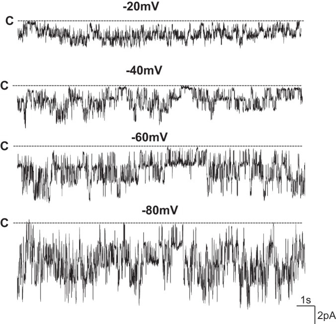
Fig. 3.

Channel recording shows the K channel activity at different holding potentials in the primary cultured mouse corneal epithelial cells. The experiment was perforated in a cell-attached patch (in 4 out of 12 patches) with 140 mM NaCl/5 mM KCl in the bath and 140 mM KCl in the pipette. The cell membrane holding potentials are indicated on the top of each trace and the channel close state was indicated by a dotted line and “C”.
