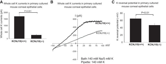
Fig. 5.
A: bar graph summarizes the results of experiments in which Ba2+-sensitive K currents were measured with perforated whole cell recording technique at −60 mV. The pipette solution and the bath solution contained a symmetrical 140 mM KCl (n = 5). B: whole cell patch recording shows the whole cell currents with 140 mM NaCl/5 mM KCl in the bath and 140 mM KCl in the pipette in the primary culture corneal epithelial cells from WT (Kcnj10+/+) and Kcnj10−/−, respectively. The K+ reversal potential was determined by the potential at which the whole cell currents changed from inward (negative) to outward (positive) (n = 4). C: bar graph summarizes the results in which K+ reversal potentials were measured with the perforated whole cell recording with 140 mM NaCl/5 mM KCl in the bath and 140 mM KCl in the pipette (n = 4).

