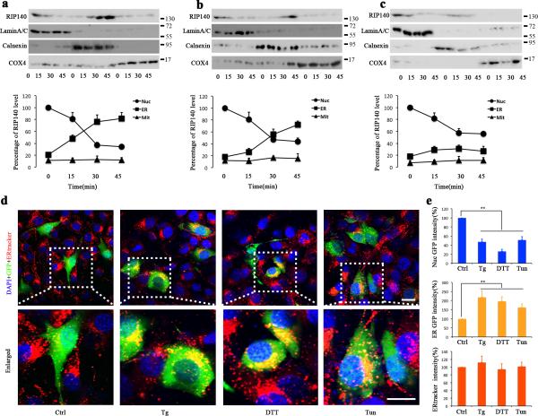
Figure 2. UPR induces translocation of RIP140 to the ER.
a-c, Western blot analyses of RIP140 levels in nuclear (Nuc), endoplasmic reticulum (ER) and mitochondrial (Mit) fractions of HT22 cells at different time points following treatment with 1μM thapsigargin(Tg) (panel a), 2μM dithiothreitol (DTT) (panel b) or 500 μM tunicmycine(Tm) (panel c) treatment. The relative protein level was analyzed as a percentage of lamin A/C for nuclear, calnexin for ER and COX4 for Mit. The RIP140 level in Nuc of control group was calculated as 100%. d, Representative confocal microscopy images indicating RIP140 cytoplasmic translocation and localization to ER in HT22 cells after treatment with 1 μM Tg, 2 μM DTT and 50 μM Tm. Green fluorescence indicates infected cells with Lentivirus carrying GFP-RIP140. Red fluorescence shows ER tracker. Nuclei are labeled with DAPI (blue). Scale bars, 20 μm. e, Percentage of nuclear and cytoplasmic GFP fluorescence or red fluorescence intensity from three independent experiments (43 cells were included in each group), showing RIP140 and ER intensity, respectively, in treated cells compared to control cells. Results are presented as means ± SEM.,**p<0.01 compare to control group as determined by Student's t-test.

