FIG. 5.
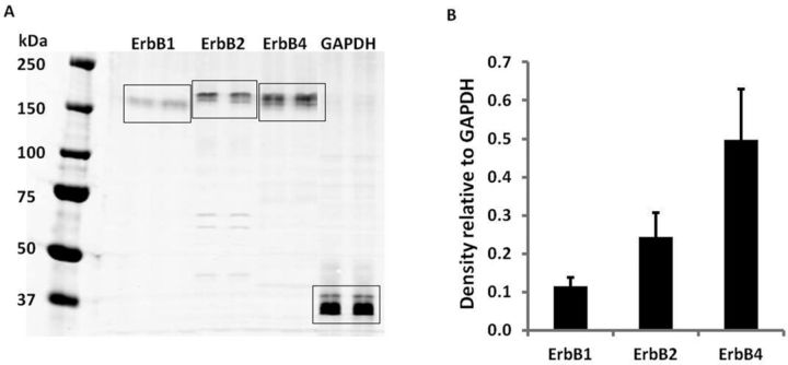
Expression of ErbB1, ErbB2, and ErbB4 receptors in hiPSC-CMs. Four-hundred microgram of total protein in 450 μl was loaded in the gel and each antibody marker was probed in duplicate lanes. GAPDH served as the loading control. (A) Representative Western blot. (B) Relative density of the bands normalized to GAPDH. Each data point represents the mean ± SE of three independent experiments.

