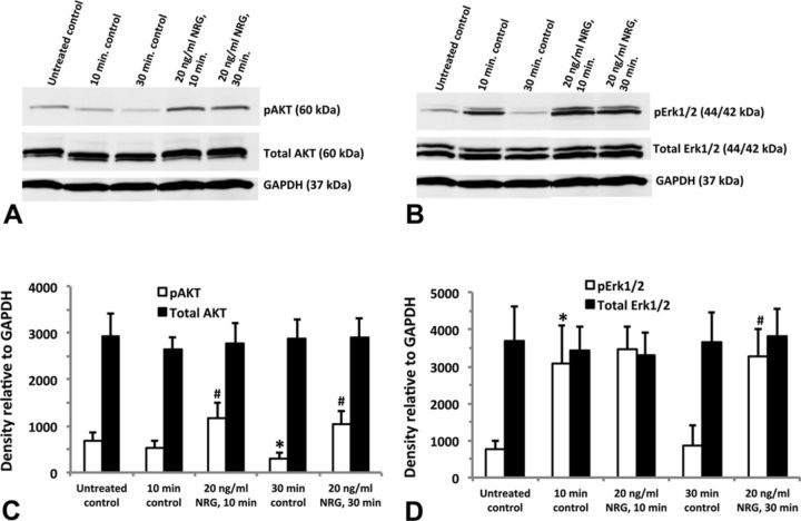
FIG. 6.
Activation of downstream ErbB signaling proteins in hiPSC-CMs. Cells treated with 20-ng/ml neuregulin-1β (NRG) for either 10 or 30 min; untreated controls did not receive fresh media, whereas the time-matched controls (at 10 or 30 min) as well as the neuregulin-treated groups did receive fresh media at the time of treatment. Representative Western blot for AKT (A) and Erk1/2 (B); 10 μg total protein was loaded into each individual well of a 10-well mini-gel (50 μl per well). Blots were first probed for phosphorylated AKT and Erk1/2, then stripped and re-probed for total AKT, total Erk1/2, and GAPDH. Relative density of the AKT bands normalized to GAPDH (C). Relative density of the Erk1/2 bands normalized to GAPDH (D). Each data point represents the mean ± SEM of three independent experiments. *#Statistically significant at p < 0.05: *pAKT 30-min control significantly less than untreated control; #pAKT 10-min and 30-min NRG treatment significantly greater than time-matched controls; *pErk1/2 10-min control significantly greater than untreated control; #pErk1/2 30-min NRG significantly greater than 30-min control.

