
Figure 3.
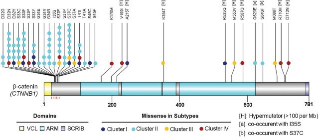
Somatic missense mutations in the CTNNB1 gene relative to the structure of the encoded protein. Schematic diagram of the β-catenin protein (accession no. NP_001091679) showing the positions of individual somatic alterations identified in endometrioid endometrial tumors. Alterations in Cluster I (dark blue), II (cyan), III (light yellow) and IV (dark red) tumors are distinguished. Alterations associated with hypermutated samples (mutation rate > 100 per Mb) are indicated by [H]. The sites of phosphorylation by GSK-3β are indicated by the red lines underneath the protein structure. Known functional domains of the protein are indicated. ARM = Armadillo/beta-catenin-like repeats; SCRIB = interaction with SCRIB (by similarity); VCL = interaction with VCL (by similarity).