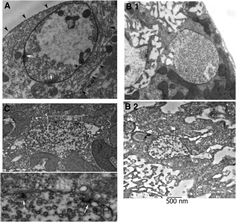
Figure 3.
Ultrastructure of new boutons. A, Micrograph of a typical mature bouton with clustered vesicles, SSR (arrowheads), and AZs (arrows). B, Images showing new boutons at in the vicinity of pre-existing clusters. Arrowheads (B.1 and B.2) show cisternae in the vicinity of the boutons in muscle tissue, which may represent a precursor of the forming SSR. C, Micrograph showing a new bouton with two AZs (T-bars) surrounded by vesicles. The boxed area is enlarged at the bottom panel. Note the absence of the SSR around the bouton, even though the T-bars (white arrows) appear to be fully formed.

