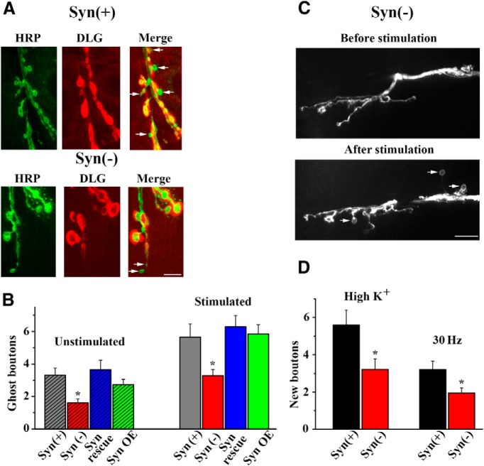
Figure 4.
Neuronal outgrowth is inhibited in the absence of synapsin. A, HRP/DLG immunolabeling of stimulated preparations shows ghost boutons (arrows) with no DLG labeling. B, Stimulation promotes the growth of ghost boutons (p < 0.0001 per 2-way ANOVA), and the number of ghost boutons is significantly reduced in Syn(−) preparations (p < 0.05). Data collected from 36, 33, 20, and 29 segments (wild type [Syn(+)], mutant [Syn(−)], rescue (Rescue), and over-expression (OE), respectively] in unstimulated preparations and 43, 35, 28, and 32 segments in stimulated preparations (>6 larvae per line per condition). C, Assessing activity-dependent formation of new boutons in Syn(−) preparations with GFP-tagged neuronal membranes using live confocal imaging. D, Activity-dependent formation of new boutons is reduced in Syn(−) preparations. Synapsin gene deletion produces a significant (p = 0.01 per 2-way ANOVA) reduction in the number of new boutons formed either upon high-K+ patterned application (high K+) or upon electrical stimulation of the nerve (30 Hz). Data collected from 42 Syn(+) and 28 Syn(−) segments with high-K+ stimulation, and 54 Syn(+) and 22 Syn(−) segments with electrical stimulation (≥6 larvae per line per condition).

