
Figure 1.
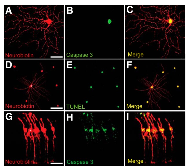
Injection of CytC into single cells results in GJ-mediated apoptosis of neighboring coupled cells. A–F, Representative photomicrographs of uncoupled (A–C) and coupled (D–F) RGCs in whole-mount retinas injected with the mixture of Neurobiotin and CytC to visualize GJ coupling and to induce cell apoptosis, respectively. C, Overlay of A and B represents apoptosis of the impaled RGC, but not any neighboring cells. F, Overlay of D and E represents that apoptosis was spread among neighboring cells coupled to impaled RGC (asterisk). G–I, Representative confocal image of coupled Müller cells in retinal vertical section, one of which (asterisk) was injected with Neurobiotin + CytC (G) and then labeled with anti-caspase 3 antibody (H) to detect apoptotic cells. I, Overlay of G and H represents spread of death in coupled neighboring Müller cells. Scale bars: A–C, G–I, 20 μm; D–F, 50 μm.