Figure 3.
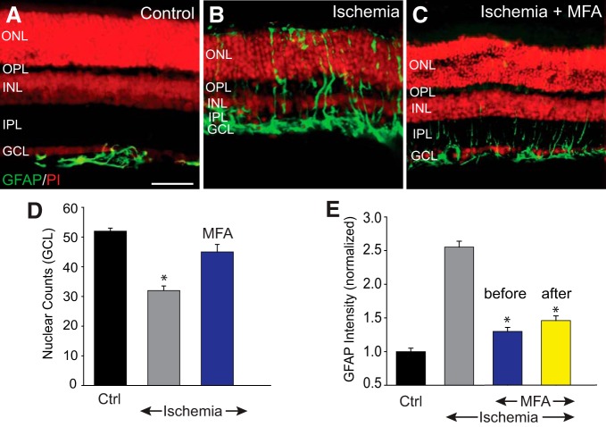
Blockade of GJs reduced retinal injury and cell death after ischemia-reperfusion. A, In control retinas, GFAP expression was confined exclusively to astrocytes in the GCL and nerve fiber layer (NFL). B, Seven days after reperfusion, the inner retinal thickness was reduced and the GFAP immunoreactivity appeared to traverse throughout the retinal layers in the Müller cell processes. C, Intravitreal injection of MFA (500 μm, 2 μl) prevented changes in the retinal morphology and maintained normal GFAP immunoreactivity. D, Histogram represents the nuclear counts in the GCL of vertical sections of control (n = 19/n = 5), and ischemic retinas in the presence (n = 16/n = 5) or absence (n = 16/n = 5) of MFA. E, Histogram represents the GFAP immunofluorescence intensity throughout the vertical section in control and ischemic retinas with or without MFA treatment. MFA was injected intravitreally either once 30 min before (blue, n = 16/n = 5), or twice at 3 and 24 h after (yellow, n = 12/n = 3) ischemic insult. Retinal sections were counterstained with propidium iodide (PI). *p < 0.01. Scale bar, 50 μm.

