Figure 5.
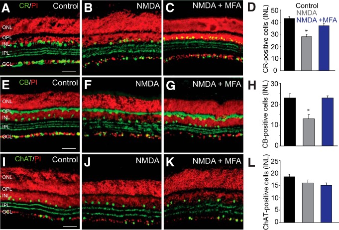
Amacrine cells that are extensively coupled to RGCs show higher susceptibility to NMDA-induced excitotoxicity. A, CR labeled a large number of cells in the INL and GCL of control retina. B, In retinas exposed for 1 h to NMDA (300 μm), the number of CR-positive cells was significantly reduced. C, In vitro treatment of retinas with MFA (50 μm, 30 min) prevented the reduction in the number of CR-IR cells in the INL. D, Histogram summarizing NMDA-induced changes in the number of CR-positive amacrine cells in the INL of untreated (n = 18/n = 3) and MFA-treated retinas (n = 10/n = 3). E, CB-labeled horizontal cells in control retina. F, Exposure to NMDA reduced the number of CB-IR cells in the proximal INL. G, Blockade of GJs with MFA prevented such reduction. H, Histogram summarizing NMDA-induced changes in the number of CB-positive amacrine cells in the INL of untreated (n = 22/n = 3) and MFA-treated retinas (n = 6/n = 3). I–L, The number of ChAT-IR amacrine cells in control retinas. No significant change in the number of ChAT-IR amacrine cells was observed in NMDA-treated retinas compared with controls (n = 20/n = 4). Retinal sections were counterstained with propidium iodide (PI). *p < 0.01 versus control. Scale bars, 50 μm.

