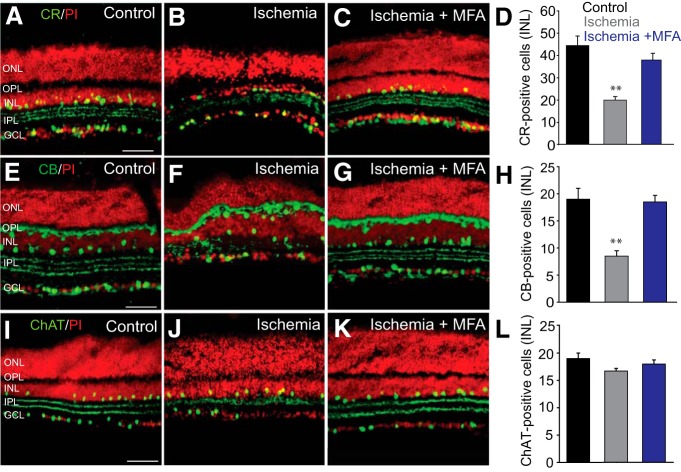
Figure 6.
Amacrine cells with a high degree of coupling to RGCs show higher susceptibility to ischemia-reperfusion injury. A–C, CR-IR cells in control retinas and in those subjected to ischemia-reperfusion injury in the absence and presence of MFA. D, Histogram summarizing alteration in the number of CR-IR amacrine cells in the INL of ischemic retinas with (n = 26/n = 5) or without (n = 48/n = 5) MFA treatment. E, CB immunoreactivity was localized to the horizontal cells and amacrine cells in the INL and sparse GCs. Three strata in the IPL were labeled as well. F, Changes in retinal morphology and the number of CB-IR cells after ischemia/reperfusion. G, Blockade of GJs by MFA largely prevented a reduction in the number of CB-IR cells. H, Histogram summarizing changes in CB-IR cells in the INL of control and ischemic retinas untreated (n = 17/n = 3) or treated (n = 21/n = 3) with MFA. I–L, No detectable change in the number of ChAT-IR amacrine cells was observed in ischemic retinas compared with levels in controls (n = 25/n = 4 each, p > 0.1). Retinal sections were counterstained with propidium iodide. **p < 0.001 versus control. Scale bars, 50 μm.

