
Figure 2.
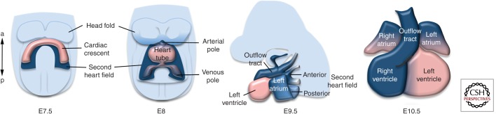
The contribution of the second heart field to heart tube extension. Cartoon showing the progressive addition of second heart field progenitor cells (dark blue) to the elongating heart tube between 7.5 and 9.5 d of mouse development. In the midgestation heart (right), second heart field–derived parts of the heart are indicated in blue. (From Kelly 2012; reproduced, with permission.)