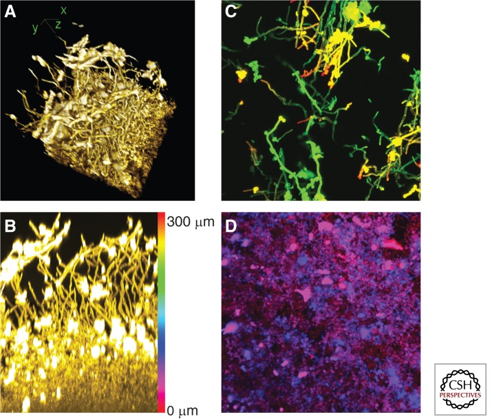
Figure 1.
Four views of a C. albicans biofilm. These images portray a single biofilm grown in yeast-peptone-dextrose medium at 37°C. It was embedded and stained with Alexafluor 594-conjugated Concanavalin A. Each image processes a portion of a confocal laser scanning microscope dataset with National Institutes of Health ImageJ software. (A) Three-dimensional reconstruction of the biofilm sample. Hyphae at the top of the biofilm are readily visible. x-, y-, and z-axes are diagrammed. (B) Side projection view of the x–z plane. Approximately 25% of the x-axis from the sample was used, permitting visualization of hyphae in the upper portion of the biofilm. Brightly stained aggregates are extracellular matrix material. The color scale bar represents the depth of the biofilm, 300 μm, and indicates the pseudocolor scale used for apical projections in C and D. (C) Apical projection of upper (substrate-distal) 100-μm region. This image shows the x–y plane. Hyphae extend from 200 μm from the substrate (green) to 300 μm from the substrate (red). Some yeast cells and extracellular material are apparent. (D) Apical projection of basal (substrate-proximal) 100-μm region. This image once again shows the x–y plane. Yeast cells are evident from the substrate level (red) to 100 μm above the substrate (blue), although light penetration limits the resolution of the image. Amorphous extracellular material is also evident.

