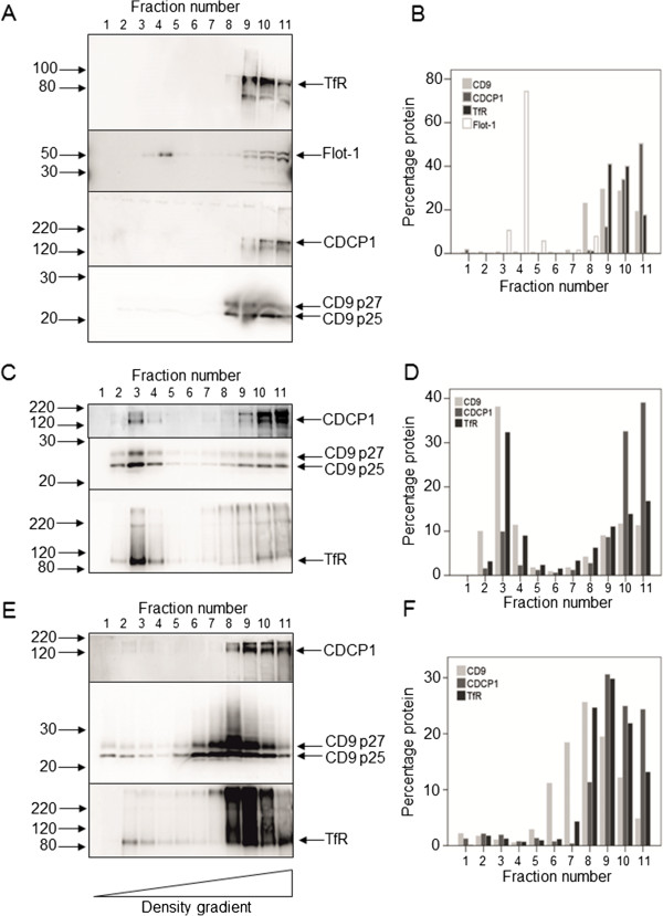
Figure 3.

Analysis of CDCP1 and CD9 in detergent-resistant membrane (DRM) fractions of HT-29 colon cancer cells. HT-29 cells were plated at 3.8 × 10⁶ cells per T175 flask and grown to confluence. The cells were scraped into MBS containing either 0.5% Triton-X100, Brij58 or Brij97 on ice. The lysates were fractionated by sucrose density gradient centrifugation as described in Materials and Methods. Eleven 0.5 ml fractions were collected, with fraction 1 being the top and fraction 11 the bottom of the gradient. The fractions were analysed by Western blotting using the following antibodies (final concentrations in parentheses): CDCP1 (1 μg/ml goat ab1377), CD9 (2 μg/ml mouse monoclonal 602–29), TfR (100 ng/ml mouse monoclonal H68.4 (13–6800)), Flotillin-1 (50 ng/ml). A, B: Triton X-100 extraction; C, D: Brij58 extraction; E, F: Brij97 extraction. B, D, F: Quantitation of the Western blots shown in panels A,C and E respectively using Advanced Image Data Analyzer (AIDA) software.
