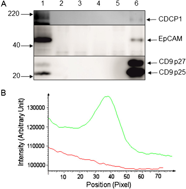
Figure 4.

Co-immunoprecipitation of CDCP1 and CD9. SW480 cells were lysed in 1% (w/v) Brij97, 10 mM Tris–HCl (pH 7.4), 150 mM NaCl, 1 mM CaCl2, 1 mM MgCl2. A. Immunoprecipitation from whole cell extracts of SW480 cells was performed using 5 μg of ALB6 anti-CD9 monoclonal antibody (lane 6) or isotype-matched control antibody (lane 5). Immunoprecipitates were analysed by Western blotting with antibodies against either CDCP1 (ab1377), top panel; EpCAM (ab32392), middle panel or CD9 (602.29), lower panel. Further controls for the specificity of immunoprecipitation and Western blotting are: lane 1: 15 μg whole cell extract, lanes 2 and 3: 5 μg of isotype-control antibody and anti-CD9 antibody respectively; lane 4: SW480 proteins bound to protein G beads. B. Densitometric analysis of the Western blot for CDCP1. A portion of the blot corresponding to the CDCP1 p135 region was analysed in the tracks generated by immunoprecipitation with the control (red) and anti-CD9 (green) antibodies using AIDA software.
