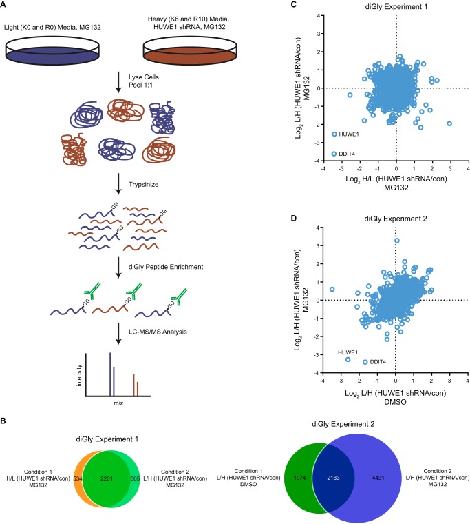
FIGURE 2.
Quantitative diGly proteomics implicates DDIT4 as an HUWE1 substrate. A, schematic illustrating a quantitative diGly proteomics experiment to identify possible HUWE1 substrates. B, Venn diagram of all diGly peptides identified and quantitated in the two quantitative diGly proteomics experiments. C and D, scatter plot representing peptides identified by LC-MS/MS following diGly affinity purification in BT-549 cells. C, log2 ratios of H/L (HUWE1 shRNA/con) on the x axis and L/H (HUWE1 shRNA/con) conditions on the y axis from cells treated with MG132. D, log2 ratios of L/H (HUWE1 shRNA/con) from cells treated with DMSO on the x axis and cells treated with MG132 on the y axis. con, control, L, light, and H, heavy.

