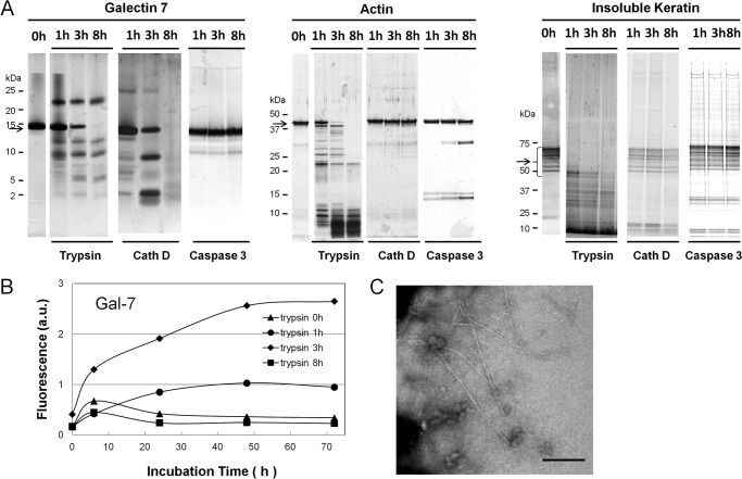
FIGURE 7.
Detection of amyloidogenic peptide in proteolytic fragments of Gal-7, actin, and cytokeratins. A, proteolytic patterns of Gal-7, actin, and insoluble cytokeratins with trypsin, cathepsin D (Cath D) and caspase 3. Gal-7 (200 μg) (left), actin (200 μg) (middle) and insoluble cytokeratins (300 μg) (right panel) were digested with trypsin, cath D, or caspase 3 at room temperature or 37 °C for 1, 3, and 8 h at their optimal pH. The digests were resolved on SDS-PAGE and then visualized with silver stain. Arrows on the left side of each panel indicate the migration positions of Gal-7, actin, and cytokeratins, respectively. B, ThT fluorescence of trypsin digests of Gal-7. Aliquots of 0, 1, 3, and 8 h-tryptic digests were incubated in 100 mm citrate buffer, pH 2.0, containing 100 mm NaCl at 37 °C for 0, 6, 24, 48, and 72 h. C, electron microscopic observation of tryptic digests of Gal-7. Aliquot of 3 h trypsin digests after 72 h incubation was taken and subjected to electron microscopic analysis. Bar; 200 nm.

