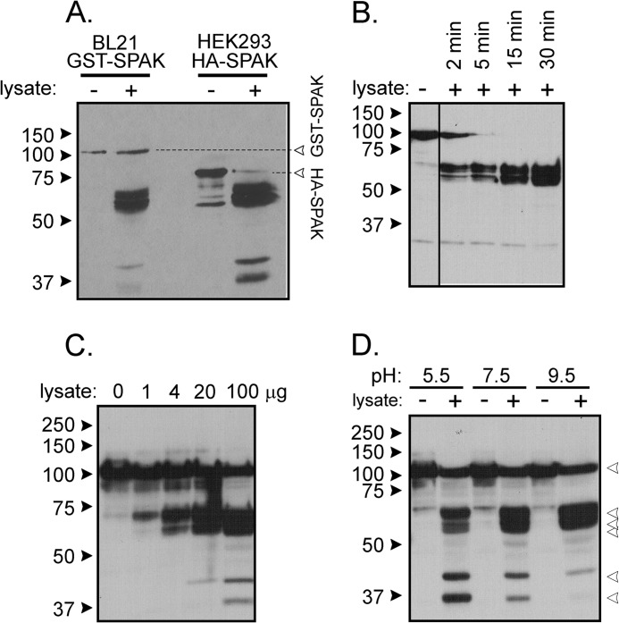
FIGURE 3.
Properties of kidney lysate cleavage of SPAK fusion protein. A, incubation of GST-SPAK isolated from bacteria and HA-SPAK isolated from HEK293 cells with kidney lysate (2 μl) cells results in proteolytic cleavage independent of the nature of the added 5′ sequence. B, incubation of GST-SPAK with kidney lysate (2 μl) for short incubation times shows time dependence. C, incubation of GST-SPAK with increasing amounts of kidney lysate shows dose dependence. D, reaction between GST-SPAK and kidney lysate (2 μl) at three different pHs. The pH was adjusted to 7.5 at the end of the reaction before protein denaturation and gel loading.

