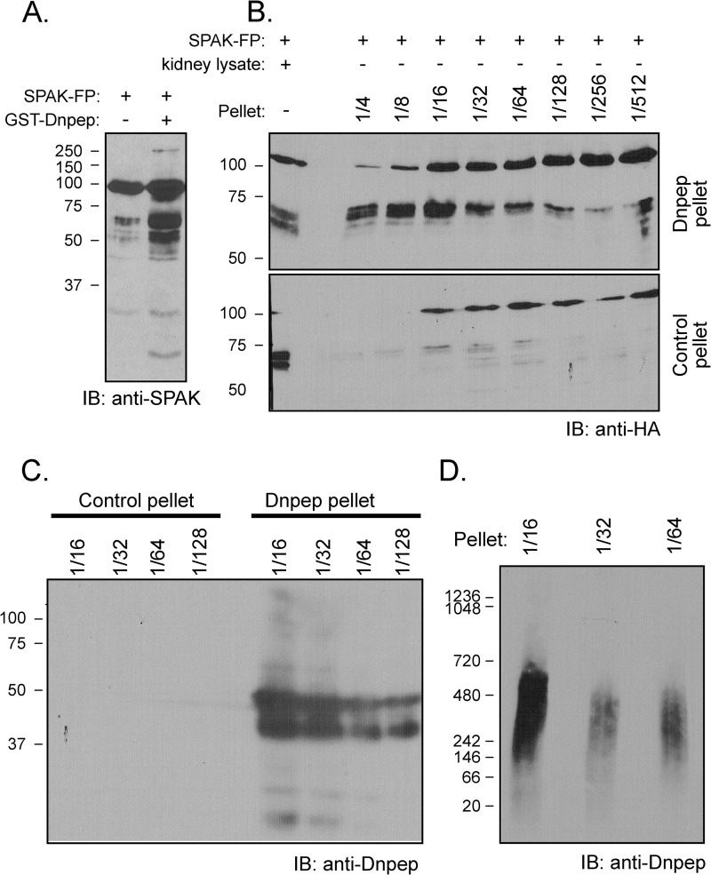
FIGURE 8.
Dnpep fusion protein cleaves SPAK fusion protein. A, reaction between GST-SPAK and GST-Dnpep. GST-Dnpep fusion protein (FP) was detected in the supernatant of lysed Dnpep-transformed bacteria and purified by a glutathione-Sepharose column. SPAK fusion protein (2 μl) was incubated (or not) in HEPES buffer with 10 μl of GST-Dnpep fusion protein for 1 h at 37 °C. IB, immunoblot. B, reaction between GST-SPAK and His-Dnpep. His-Dnpep fusion protein was detected in inclusion bodies in the pellet of lysed Dnpep-transformed bacteria. Different dilutions (4–512×) of pellet resuspended in lysis buffer (38 μl) were incubated with 2 μl of SPAK fusion protein for 1 h at 37 °C. The reaction between 2 μl of kidney lysate and 2 μl of SPAK fusion protein was used as positive control. The reaction between the pellet of GST-unspecific fusion protein and SPAK fusion protein was used as negative control. C, detection of Dnpep in SDS-PAGE Gel. Dnpep was detected in the pellet of Dnpep-transformed bacteria whereas it was absent in the pellet of unspecific protein-transformed bacteria. D, detection of Dnpep in NativePAGE 3–12% Bis-Tris gel. Dnpep was detected in the pellet of Dnpep-transformed bacteria.

