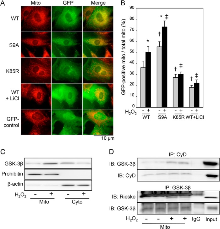
FIGURE 1.
Kinase activity-dependent mitochondrial translocation of GSK-3β under the condition of oxidative stress. A, fluorescence images obtained from time lapse observation. H9c2 cells were transfected with GFP-tagged GSK-3β (WT), constitutively active mutant GSK-3β (S9A), kinase-inactive mutant GSK-3β (K85R), or GFP alone (GFP control) and then stained with MitoTracker Red. Photos at 3 min after exposure of cells to H2O2 (10 μmol/liter) in the presence or absence of LiCl (30 mmol/liter) are shown. B, quantification of mitochondrial (mito) localization of each plasmid. MitoTracker-stained area overlapped with GFP signal was expressed as a percentage of total MitoTracker-stained area. *, p < 0.05 versus cells transfected with the same plasmid and treated with a vehicle; †, p < 0.05 versus cells transfected with WT and treated with a vehicle; ‡, p < 0.05 versus cells transfected with WT and exposed to H2O2. C, representative immunoblotting for total GSK-3β in mitochondrial fractions (Mito) and cytosolic fractions (Cyto). Prohibitin and β-actin were used as markers of the mitochondria and cytosol, respectively. Three separate experiments showed similar results. D, interaction of GSK-3β with mitochondrial proteins. Immunoblots (IB) for GSK-3β co-immunoprecipitated (IP) with cyclophilin D (CyD) and Rieske co-immunoprecipitated with GSK-3β with or without exposure to H2O2 (100 μmol/liter; 4 h) are shown. Error bars represent S.E.

