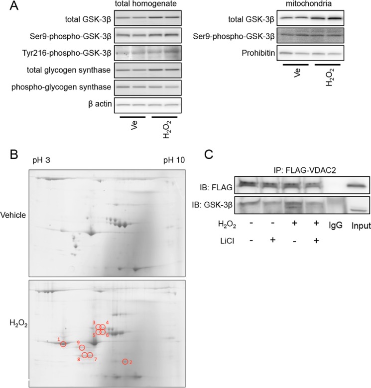
FIGURE 4.
Effects of H2O2 on GSK-3β activity and its interaction with other proteins. A, H9c2 cells were exposed to 100 μmol/liter H2O2 or a vehicle (Ve) for 4 h and harvested. Results of Western blotting for total GSK-3β, Ser-9-phospho-GSK-3β, Tyr-216-phospho-GSK-3β, total glycogen synthase, and phospho-glycogen synthase in the total homogenate (left panels) and total GSK-3β and Ser-9-phospho-GSK-3β in the mitochondrial fraction (right panels) are shown. β-Actin and prohibitin serve as loading controls. B, representative two-dimensional gels stained by Coomassie Blue. Samples were obtained from GSK-3β immunoprecipitates of H9c2 cells that were treated with a vehicle or H202 (100 μmol/liter) for 3 h. Results of four experiments for vehicle-treated cells and four experiments for H2O2-exposed cells were similar. Spots that exhibited increases in the density by 3-fold or more after exposure to H2O2 are labeled as 1–9. C, HEK293 cells transfected with FLAG-VDAC2 were exposed to H2O2 (100 μmol/liter; 4 h) in the presence or absence of LiCl (30 mmol/liter). Immunoblots (IB) for FLAG and GSK-3β co-immunoprecipitated (IP) with FLAG-VDAC2 are shown.

