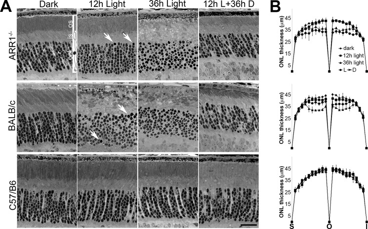
FIGURE 1.
Light exposure causes rapid photoreceptor cell death in ARR1−/− and BALB/c mice. 1-month-old ARR1−/−, BALB/c, and C57/B6 mice were exposed to 5000-lux white light, pupils undilated, for 12 (12h Light) or 36 h (36h Light). Another group was dark adapted for 36 h after 12 h of light exposure (12h L+36h D). A, retinal morphology of the indicated mice before and after light exposure. Arrows point to pyknotic nuclei. No changes in retinal morphology were detected in C57/B6 mice. Retinal layers are depicted by the rod photoreceptor cell diagram: os, outer segment; is, inner segment; onl, outer nuclear layer. Scale bar, 20 μm. B, morphometric measurements of outer nuclear layer thickness were taken at 20 equal distance positions from superior (S) to inferior (I) poles of the retina. Each point represents mean ± S.D. (n ≥ 3 independent mice) per group. We performed ANOVA for ONL thickness in the central region, which showed significant p values for the 36-h light (p < 0.0009) and 12-h light up to the 36-h dark time points (p < 0.002). These were followed up by pairwise comparisons using t test. For the 36-h light time point, both ARR1−/− and BALB/c were significantly different from C57 (p < 0.006 and p < 0.004, respectively), whereas values for ONL thickness for ARR1−/− and BALB/c were not statistically different. For 12-h light to 36-h dark condition, the p values are as follows: C57 versus ARR1−/−, p < 0.004; C57 versus BALB/c, p < 0.04; and ARR1−/− versus BALB/c, p < 0.02.

