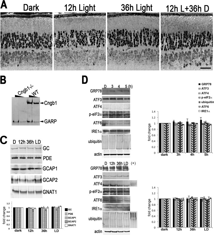
FIGURE 8.
Absence of light damage in the Cngb1−/− retina. A, retinal morphology of Cngb1−/− mice before and after the light damage protocol. Scale bar, 20 μm. B, Western blot of whole retinal homogenate prepared from Cngb1−/− and WT mice. Two different concentrations were loaded per sample. The blot was probed with monoclonal antibody 4B1 made against the GARP epitope. C, Western blots of whole retinal homogenates prepared from Cngb1−/− mice following light damage protocol probed for the indicated phototransduction proteins. The signals were quantified and plotted in the bar graph below (means ± S.E., n ≥ 3). No significant differences were detected by ANOVA. D, Western blots of the retinal homogenates probed with the indicated UPR markers. ARR1−/− retinal homogenates from 3-h light-exposed mice were included as positive control (+) for some lanes. The signals were quantified and plotted in the bar graph (means ± S.E., n ≥ 3). No significant differences were detected by ANOVA. D, dark adapted; LD, 12 h light exposed followed by 36 h dark adaptation.

