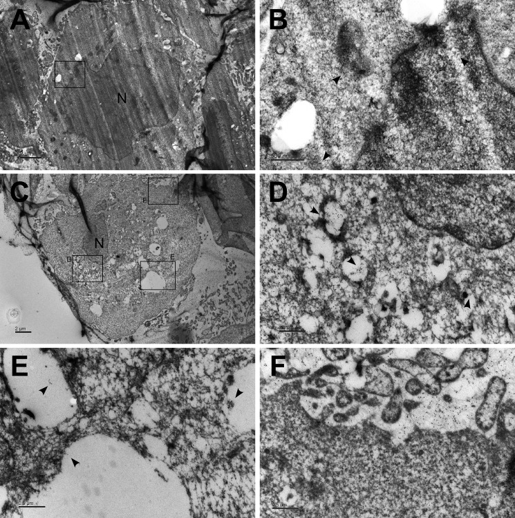
FIGURE 4.
GFP-ASC localizes to autophagic vacuoles following AIM2 inflammasome activation. A, NPC TW02 cells were transfected with GFP-ASC for 48 h and then treated with/without poly(dA-dT). Ultrathin sections were stained with gold particles targeting the anti-GFP antibodies and observed by electron microscopy. Scale bar, 2 μm. B, image magnified from A. Scale bar, 500 nm. C, NPC TW02 cells were transfected with GFP-ASC for 48 h, treated with poly(dA-dT) for 4 h, and then examined as described in A. Scale bar, 2 μm. D–F, images magnified from C. Scale bar, 500 nm. The arrowhead indicates the gold particles targeting GFP-ASC. N indicates the nucleus.

