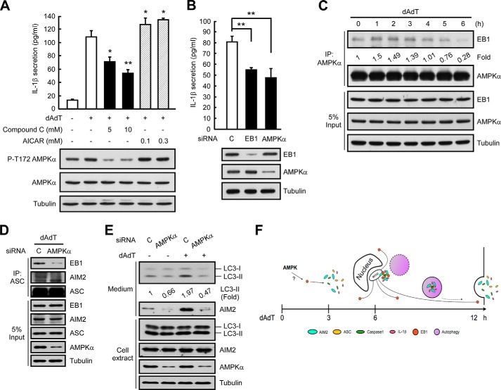
FIGURE 7.
AMPK may regulate EB1. A, HK1 cells were pretreated with the indicated doses of compound C or AICAR for 6 h, and then treated with poly(dA-dT) for 12 h. Culture media and cell extracts were subjected to IL-1β ELISA and Western blot analysis, respectively. *, p < 0.05; **, p < 0.01. B, HK1 cells were pretreated with control siRNA or AMPKα siRNA for 48 h and then treated with poly(dA-dT) for 12 h. Culture media and cell extracts were subjected to IL-1β ELISA and Western blot analysis. *, p < 0.05; **, p < 0.01. C, NPC TW02 cells were treated with poly(dA-dT) for the indicated durations, and cell extracts were subjected to immunoprecipitation. D, NPC TW02 cells were treated with control siRNA or AMPKα siRNA for 48 h, treated with poly(dA-dT) for 6 h, extracted, and subjected to immunoprecipitation. E, NPC TW02 cells were treated with control siRNA or AMPKα siRNA for 48 h and then treated with/without poly(dA-dT) for 12 h. Culture media and cell extracts were subjected to Western blot analysis. F, the time course of poly(dA-dT) treatment-induced EB1-mediated AIM2 inflammasome delivery via autophagic vacuole shedding, which occurs as follows: 1) AMPK regulates EB1; 2) EB1 regulates the MTOC localization of the AIM2 inflammasome speck; and 3) EB1 elicits AIM2 inflammasome release via autophagic vacuole shedding.

