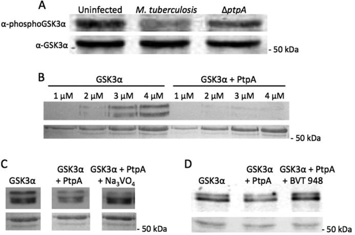
FIGURE 4.

Western blot analyses of PtpA dephosphorylation of GSK3α in vivo and in vitro. A, THP-1 cells were infected with M. tuberculosis or ΔptpA M. tuberculosis. Cellular extracts were harvested 18 h post-infection, and 50 μg of it was used for Western blotting in which the anti-phospho-GSK3α (Tyr(P)279) antibody was utilized. The bottom panel represents the membrane probed with anti-GSK3α. The molecular mass of GSK3α is 50.981 kDa. B, different concentrations of GSK3α (1–4 μm) with and without PtpA (0.04 μm) were incubated and developed by enhanced chemiluminescence. The bottom panel represents the Ponceau-stained membrane showing equal loading of samples. C and D, fixed concentration of GSK3α (3 μm) with and without PtpA (0.04 μm) was incubated with the tyrosine phosphatase inhibitor Na3VO4 (1.5 mm) or BVT 948 (5 μm) and developed by enhanced chemiluminescence. The bottom panels represent the Ponceau-stained membranes showing equal loading of samples.
