FIGURE 7.

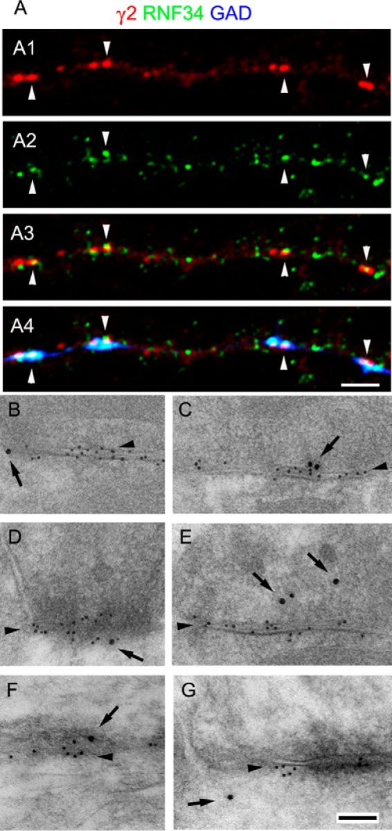
GABAergic synapses frequently have RNF34 associated with them. A, triple-labeled immunofluorescence of 21 DIV cultured hippocampal neurons with GP anti-γ2 GABAAR subunit (red, A1), Rb anti-RNF34 (green, A2), and sheep anti-GAD (blue) antibodies. A3 shows the overlay of γ2 and RNF34 fluorescence, whereas A4 shows the overlay of the three fluorescence channels. Arrowheads indicate GABAergic synapses (γ2+ and GAD+) that have co-localizing RNF34 clusters. Scale bar, 2.5 μm (A1–A4). B–G, postembedding EM immunogold double-labeling of rat brain cerebellum (B, C, and E) and cerebral cortex (D, F, and G) with Rb anti-RNF34 and mouse anti-β2/3 GABAAR subunits. The β2/3 immunolabeling (smaller gold particles) is indicated by arrowheads. GABAergic synapses are identified by their morphology and the concentration of β2/3 GABAAR subunits. The RNF34 immunolabeling (larger gold particles, arrows) is localized at or near GABAergic synapses (B–G), GABAergic presynaptic terminals (C, E, and F), and GABAergic postsynapses (B, D, and G). The goat anti-rabbit IgG and goat anti-mouse IgG secondary antibodies were conjugated to 18 and 10 nm diameter colloidal gold particles, respectively. Scale bar, 100 nm (B–G).
