Fig. 2.
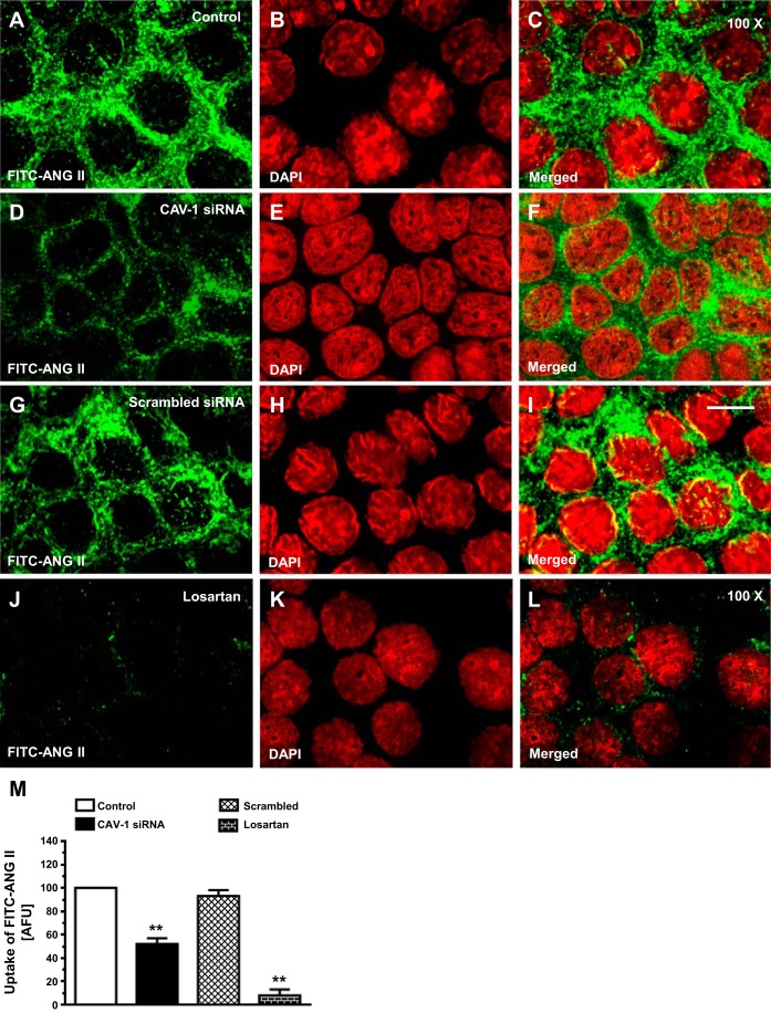
In vitro uptake of FITC-labeled ANG II in wild-type (WT) mPCT cells and the effects of CAV-1 siRNA and losartan (Los). mPCT cells were grown on glass coverslips and incubated with FITC-labeled ANG II (10 nM) at 37°C for 30–60 min. A, D, G, and J: FITC-labeled ANG II (green). B, E, H, and K: 4′,6-diamidino-2-phenylindole (DAPI)-stained images (red). C, F, I and L: merged images. M: semiquantitative ANG II uptake by mPCT cells in arbitrary fluorescence units (AFU) with control at 100. Internalized FITC-labeled ANG II was seen in the cytoplasm and perinucler regions of the cells. The FITC-labeled ANG II uptake response was attenuated by CAV-1 siRNA but not by a scrambled version of siRNA. **P < 0.01 vs. control mPCT cells. Magnification: ×100.

