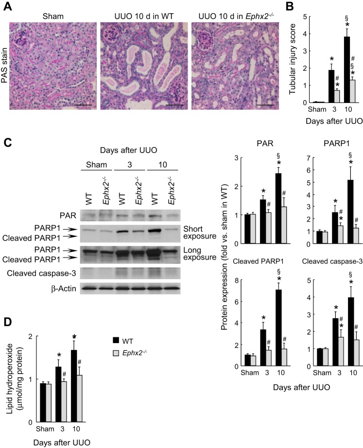
Fig. 8.
Loss of sEH suppresses tubular cell damage during interstitial fibrogenesis. A: periodic acid-Schiff (PAS) stain on kidney sections in WT or sEH-KO mice at 10 days after sham operation or UUO. Scale bars = 50 μm. B: tubular injury score represented by PAS stain in the kidneys. C: protein expression of poly ADP-ribose (PAR), poly(ADP-ribose) polymerase 1 (PARP1), cleaved PARP1, and cleaved caspase-3 in the kidneys using Western blot. The bands were quantified using Lab Works analysis software. D: lipid peroxidation indicated by lipid hydroperoxide level in the kidneys using lipid hydroperoxide assay kit. Error bars represent SD (n = 5). *P < 0.05 vs. sham. #P < 0.05 vs. WT. §P < 0.05 vs. 3 days.

