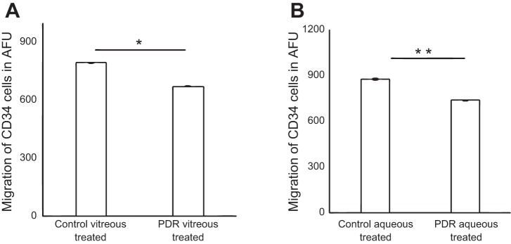
Fig. 1.

Pretreatment of CD34+ cells with proliferative diabetic retinopathy (PDR) vitreous or aqueous reduced migration of CD34+ cells. Boyden chamber assay showing migration of cells to 100 nmol/l stromal-derived factor-1. CD34+ cells treated with 5% PDR/control vitreous or aqueous and maintained at 37°C for 17 h to evaluate migration. A: migration of CD34+ cells treated with 5% PDR or control vitreous; P < 0.001. B: migration of CD34+ cells treated with 5% PDR or control aqueous (P < 0.05); x-axis represents treatment condition, and y-axis represents migration of CD34+ cells in arbitrary fluorescent units (AFU). *P = 0.0009 and **P = 0.01, significant difference compared with corresponding control. Data were analyzed by Student's t-test and represented as error bars ± SE (n = 3).
