Fig. 3.
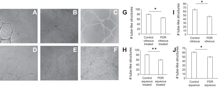
PDR vitreous and aqueous attenuate capillary tube formation. Human retinal microvascular endothelial cells were added to Matrigel-filled wells containing conditioned mixture of PDR aqueous, control aqueous, PDR vitreous, control vitreous, serum media, and VEGF (50 ng/ml); representative fields are shown. A and B: supernatants of control and PDR vitreous-treated cells. C: serum media. D and E: supernatants of control and PDR aqueous-treated cells. F: VEGF-treated cells. G–J: quantitative evaluation of tube-like structures; x-axis represents treatment condition and y-axis the no. of tube-like structures. *P = 0.04 and **P = 0.01, significant difference compared with corresponding control. Data were analyzed by Student's t-test and represented as error bars ± SE (n = 3).

