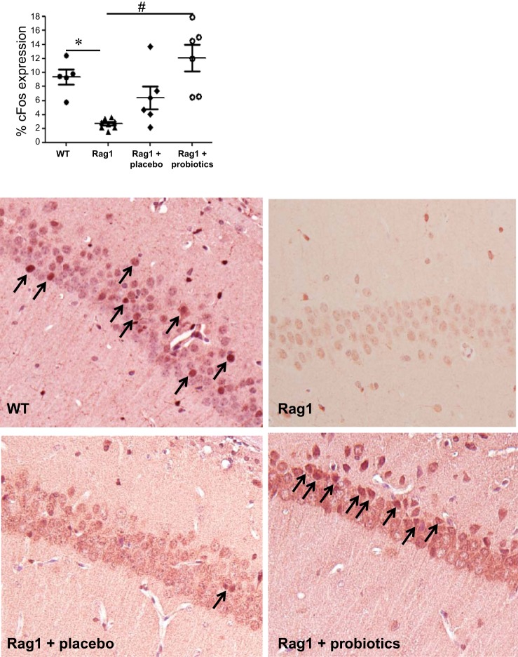
Fig. 6.
c-Fos expression in the CA1 region of the hippocampus is reduced in immunodeficient mice. Immunohistochemistry was used to assess the number of c-Fos-positive neurons in the CA1 region of the hippocampus in WT C57BL/6 and Rag1−/− mice as well as in Rag1−/− mice administered either probiotics or placebo. Means ± SE; *P < 0.05 compared with WT, #P < 0.05 compared with Rag1−/−, 1-way ANOVA, Newman-Keuls post hoc n = 6–7 mice/group. Representative images are presented in the lower panels. c-Fos-positive cells are indicated by arrows.

