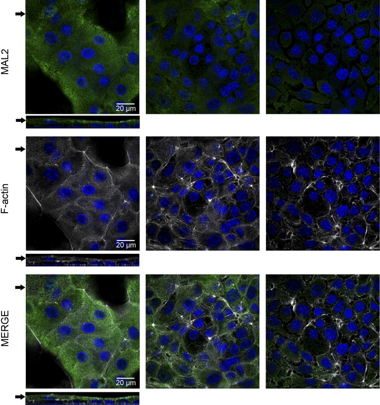
Fig. 8.
Apical polarization of ImSPEM cells. ImSPEM cells were cultured on collagen-coated Transwells at 39°C for 1 wk. Cells were immunolabeled with MAL2 (green) and F-actin (phalloidin-white). Expression patterns were analyzed by using z-stacks of the Transwells. Serial images (0.5 μm thick) taken 3 μm apart are shown. Arrows in top panels denote z-axis location (bottom panels) whereas arrows in z-axis indicate the serial images shown at top. MAL2 was localized to the subapical region of ImSPEM cells. In the bilayer of cells, MAL2 was only detected in the cells of the top layer. DAPI (blue). Scale bar = 20 μm.

