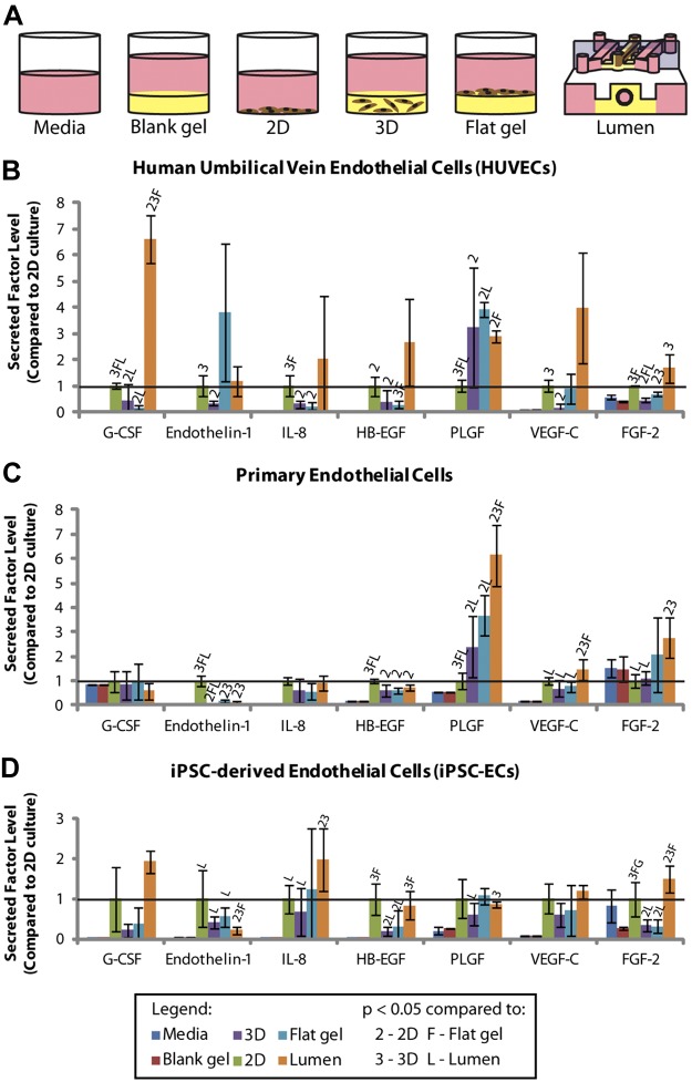
Figure 2.
A) Schematic image of culture conditions that were compared using the MagPix system. B) Secreted factor levels detected in HUVEC-conditioned medium. C) Secreted factor levels detected in iPSC-EC-conditioned medium. D) Secreted factor levels detected in primary endothelial cell-conditioned medium. FGF-2, fibroblast growth factor-2; G-CSF, granulocyte-colony stimulating factor; HB-EGF, heparin-binding-endothelial growth factor-like growth factor; IL-8, interleukin-8; PLGF, placental growth factor; VEGF-C, vascular endothelial growth factor C. Error bars= sd. Solid horizontal line represents the 2D culture condition, which is set to 1. Statistical significance between conditions was determined as a value of P < 0.05 using a Student's t test. This is denoted in each graph by a letter or number above the bar, corresponding to the comparative condition indicated in the legend.

