Fig. 4.
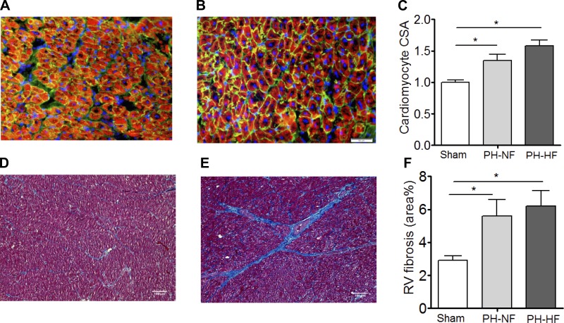
Cardiomyocyte hypertrophy and RV fibrosis. RV cardiomyocyte cross-sectional area (CSA) was examined by staining with wheat germ agglutinin to evaluate RV sections for cardiomyocyte hypertrophy. Representative sections are shown for sham-operated (A) and PH (B) pigs, with quantification of pooled data (C). RV sections were stained with Masson's trichrome to examine fibrosis. Representative sections are shown for sham-operated (D) and PH (E) pigs with quantification of pooled quantification (F). *P < 0.05.

