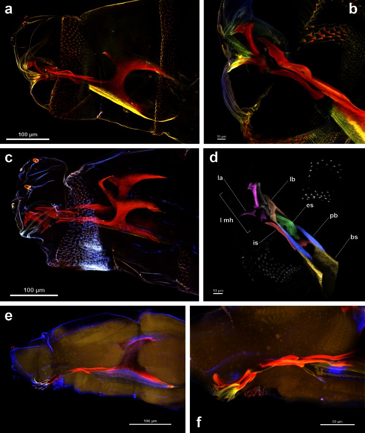
Fig. 1.
Anterior body end with the cephaloskeleton of the first instar larvae of Calliphora vicina, Lucilia sericata and Hydrotaea dentipes. a L. sericata cleared with KOH, embedded in Euparal [MIP of 28 optical sections collected with 4 lasers]. b L. sericata cleared with KOH, embedded in Euparal [MIP of 177 optical sections collected with 4 lasers]. c C. vicina cleared with KOH, embedded in water [MIP of 29 optical sections collected with 4 lasers]. d L. sericata cleared with KOH, embedded in Euparal [3D MIP of 177 optical sections same as Fig. 1b, yet rotated and collected with a 640-nm laser and visualized subsequently in B&W and with pseudo colours]. e H. dentipes cleared with Hoyer’s medium [MIP of 28 optical sections collected with 4 lasers]. f H. dentipes cleared with Hoyer’s medium [MIP of 226 optical sections collected with 4 lasers]

