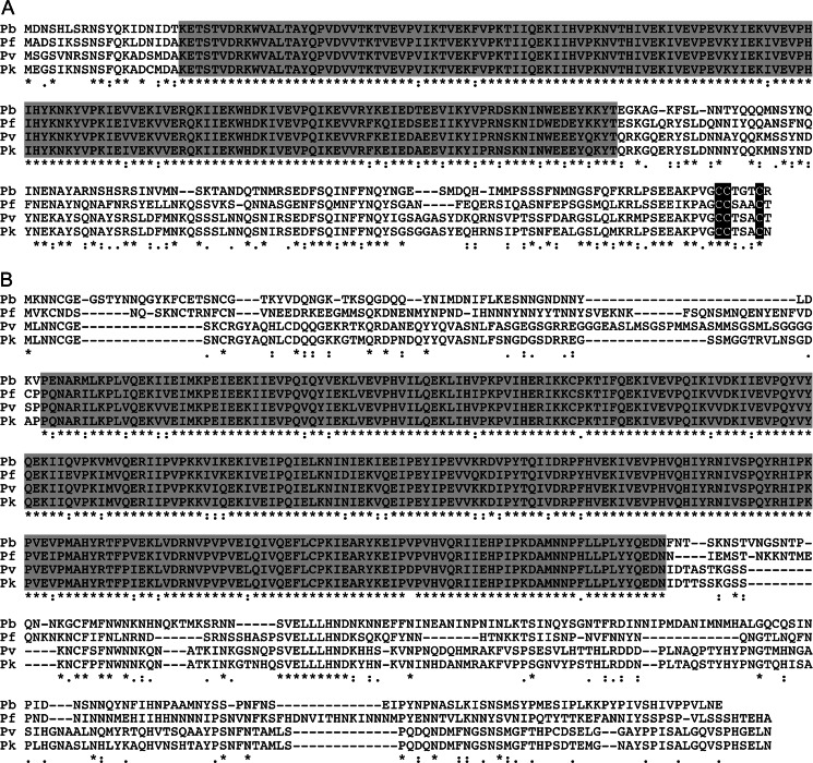
Fig. 1.
Sequence and structure of PbIMC1 proteins. Multiple amino acid sequence alignment of the predicted IMC1c (a) and IMC1e (b) proteins from P. berghei (Pb), P. knowlesi (Pk), P. vivax (Pv) and P. falciparum (Pf). Indicated are conserved alveolin domains (shaded) and gaps introduced to allow optimal alignment (hyphens). Conserved amino acid identities (asterisks) and similarities (colons and points) are indicated underneath. Also shown is a conserved cysteine motif at the carboxy-terminus of IMC1c (black shading). The alignment was made with ClustalW

