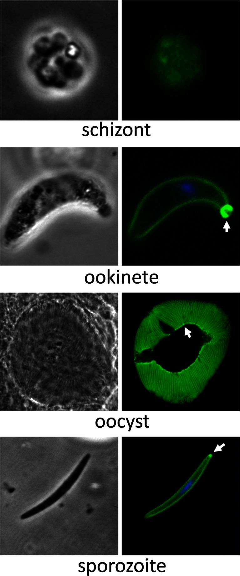
Fig. 4.
Expression and subcellular localization of PbIMC1e. Bright-field and GFP fluorescence images of schizont, ookinete, mature oocyst and sporozoite life stages. The schizont image was captured using a CCD camera due to the low levels of fluorescence, while the other images were captured by confocal microscopy. Hoechst DNA staining (blue) indicates the position of nuclei. Arrows point to posterior structures in ookinetes and sporozoites

