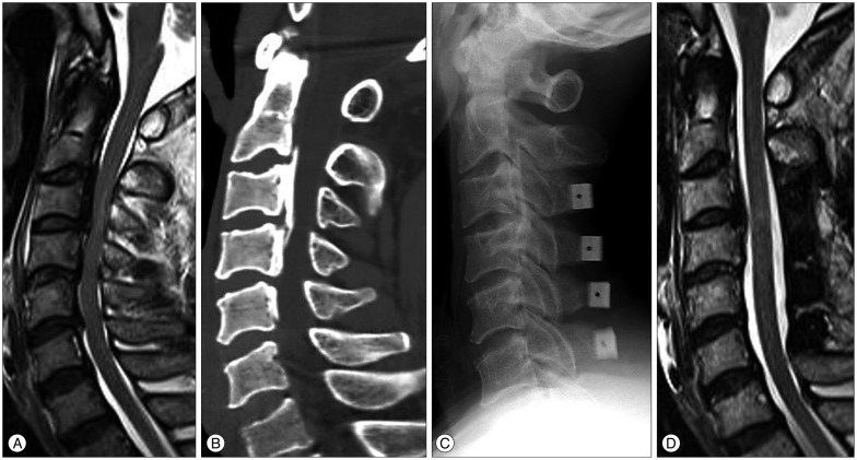
Fig. 5.
A : Preoperative T2-weighted MRI of a 61-year-old male patient with spinal cord compression at C3-4-5-6. Intramedullary high signal intensity, grade 1 was visible at the region of the marked compressed cord. His JOA score was 5. B : Cervical 3D CT showed a mixed type of ossification of posterior longitudinal ligament. Spinal canal stenosis was 46.3%. C : He underwent the cervical laminoplasty at C3-4-5-6. D : MRI of the patient from B, 12 months following the cervical laminoplasty with successful decompression of the spinal cord. The patient's Japanese Orthopedic Association score was 10. The recovery rate was 41.7%.

