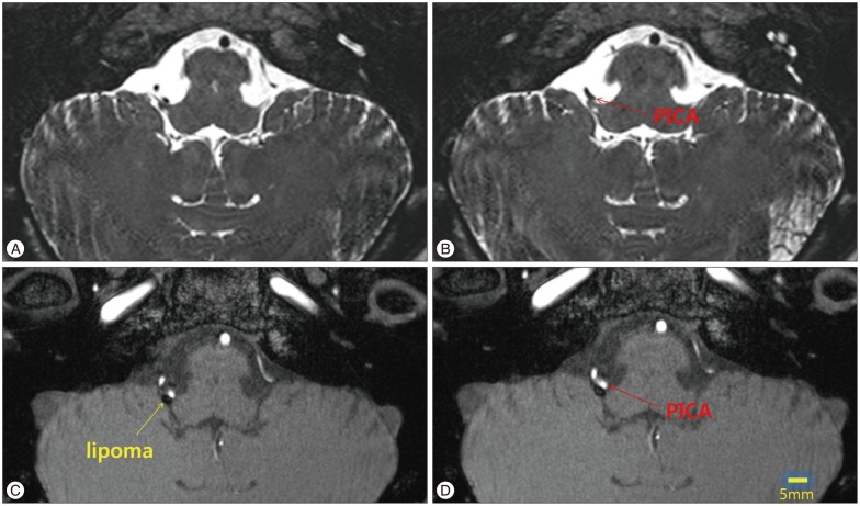
Fig. 1.
MRI finding shows a suspicious offending vessels visible at both in fast imaging employing steady-state acquisition (FIESTA) (A and B) and in time of flight (TOF) image (C and D). A small oval-shaped mass is observed as low signal intensity in TOF image (C and D). PICA : posterior inferior cerebellar artery.

